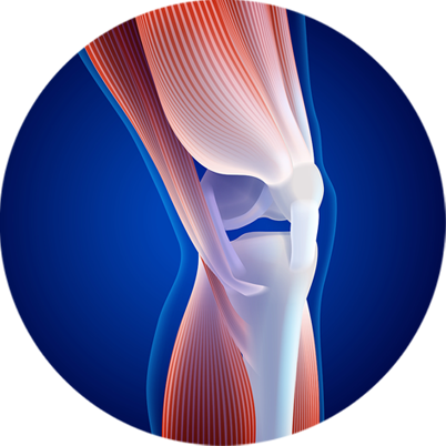
Sports knee injury refers to any injury affecting the knee joint that occurs during sport, exercise, or any athletic activities. The cartilage of the knee can be acutely injured or can gradually tear by overuse, sudden twists or impacts, or improper technique. These injuries can range from mild strains or sprains to more severe ligament tears, cartilage damage, or tendon injuries.

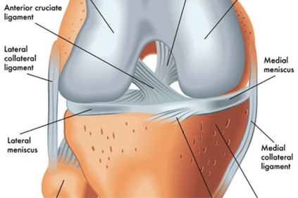
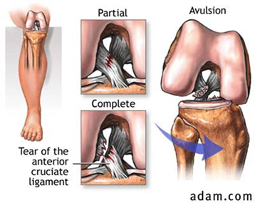
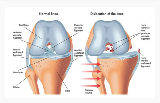
The ACL is one of the major ligaments in the knee, responsible for providing stability and preventing excessive forward movement of the tibia in relation to the femur. They occur most frequently in those who play sports involving pivoting (e.g. football, basketball, netball, soccer, European team handball, gymnastics, downhill skiing). They can range from mild (such as small tears/sprain) to severe (when the ligament is completely torn).
Causes for ACL Tear
ANTERIOR CRUCIATE LIGAMENT [ ACL] injuries can occur due to various factors, including;
Diagnosis for ACL Tear
ACL injuries are usually diagnosed based on the combination of
clinical examination, imaging studies [such as MRI], and
assessment of symptoms and mechanism of injury. Often
you may need tests to rule out other causes such as [pivot-shift the
diagnosis can be made on the basis of the physical exam alone,but
Treatment for ACL Tear
A medial collateral ligament [MCL] sprain is a common injury that affects the ligament on the inner side of the knee. The MCL play a crucial role in stabilizing the knee joint by preventing excessive side-to-side movement. A sprain occurs when the ligament is stretched beyond its normal range of motion or when it is torn partially or completely.
Common symptoms of an injury to the medial collateral ligament are:
Common Diagnosis of an injury to the medial collateral ligament are:
A doctor will typically diagnose an MCL sprain through a physical exam, which may include:
Common Treatment of an injury to the medial collateral ligament are:
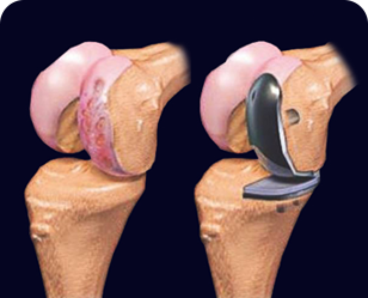
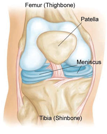
A Meniscus tear is a common injury that involves damage to the cartilage disc [menisci] located between the thigh bone [femur] and shinbone [tibia]. These C-shaped discs act as a shock absorber and help stabilize the knee joint.
Causes of Meniscus Tear
Most often, the meniscus tears during a sudden motion in which your knee twists while your foot stays planted on the ground. The tear frequently occurs while playing sports. People whose cartilage wears down (due to age or arthritis) can tear a meniscus from a motion as simple as stepping on an uneven surface. Sometimes, degeneration from arthritis causes a tear.
Types of Meniscus Tears
Meniscus tears can be classified based on their location, severity, and pattern. Common types of meniscus tears include:
Diagnosis of Meniscus Tear
Treatment of Meniscus Tear
The treatment of a meniscus tear depends on several factors including the severity of the tear, the patient’s age, activity level, and overall health. Here are some common treatment options:
Conservative Treatment:
Injection Therapy:
Surgical Treatment:
Rehabilitation:
Kneecap dislocation occurs when the round-shaped bone covering the knee (patella) moves or slides out of place. The dislocation often occurs toward the outside of the leg.
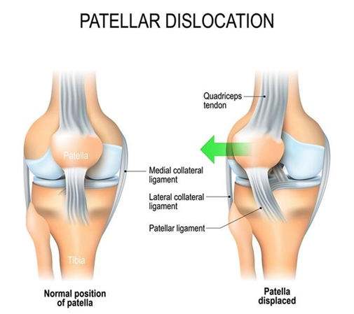
Causes of Patellar Dislocation
Symptoms of Patellar Dislocation
Typical symptoms include:
How is Patella Dislocation Diagnosed?
Healthcare providers can usually diagnose a dislocated kneecap by physically examining the knee and asking you questions about the injury. However, they’ll order radiographic imaging tests to check for any related injuries, such as torn ligaments, cartilage injury or fractures. With patellar dislocation, it is safe to correct the joint first and take pictures after.
If your dislocated patella corrected itself, you might not realize that it was dislocated. A dislocation that corrects itself is called “transient.” Afterward, your knee will still be sore and swollen, but it may look like many other more common knee injuries. In this case, imaging tests can show evidence after the fact that there was a dislocation, along with secondary injuries.
Treatment of Patellar Dislocation
Surgery:
An LCL injury is a sprain or tear to the lateral collateral ligament (LCL). The LCL is a band of tissue on the outside of your knee. It connects your thighbone to the bone of your lower leg and helps keep the knee from bending outward.
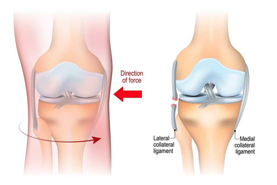
Causes of LCL Tear
LCL tears typically happen when you’re playing a sport that involves:
Diagnosis of LCL Tear
Treatment of LCL Tear
Treatment for a lateral collateral ligament (LCL) tear typically depends on the severity of the injury. Here’s an overview of potential treatment options:
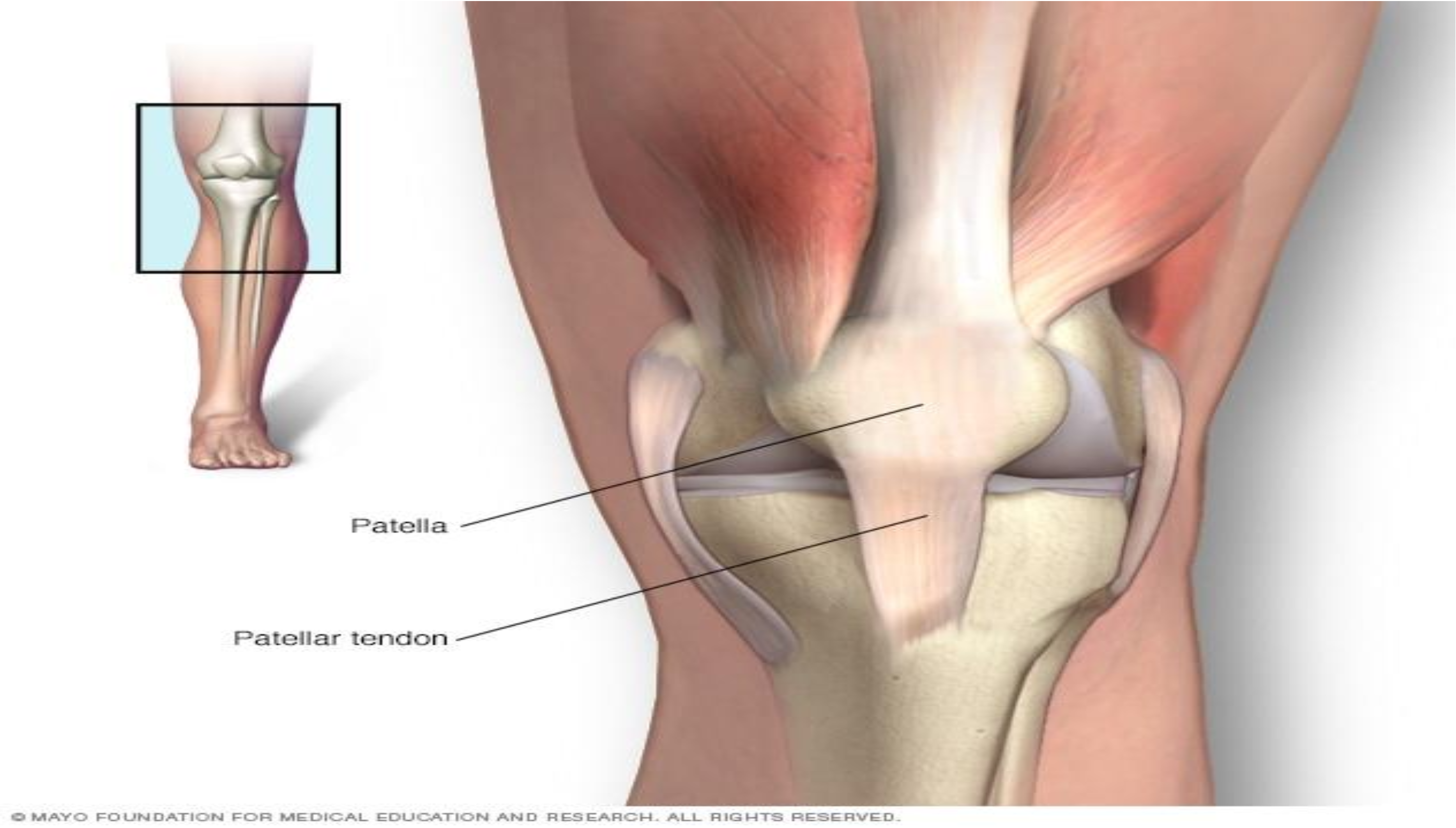
Patellar tendonitis, also known as jumper’s knee, is a common overuse of injury characterized by inflammation or degeneration of patellar tendon, which connect the kneecap[patella] to the shinbone[tibia].
Causes for Patellar Tendonitis
Patellar tendinitis is a common overuse injury, caused by repeated stress on your patellar tendon. The stress results in tiny tears in the tendon, which your body attempts to repair.
But as the tears in the tendon multiply, they cause pain from inflammation and weakening of the tendon. When this tendon damage persists for more than a few weeks, it’s called tendinopathy.
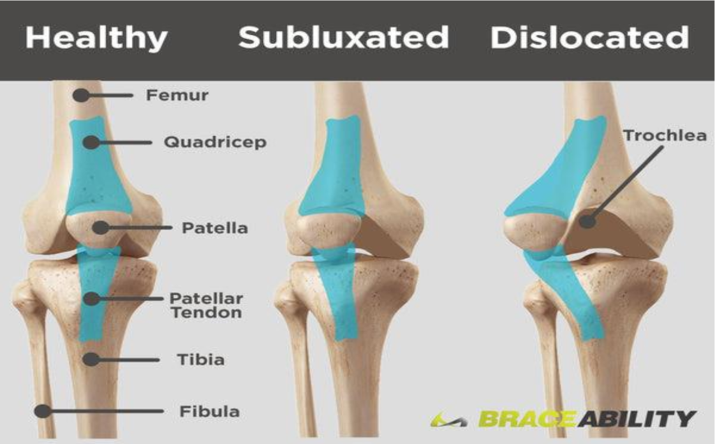
Subluxation is another word for partial dislocation of a bone. Patellar subluxation is a partial dislocation of the kneecap (patella). It’s also known as patellar instability or kneecap instability.The kneecap is a small protective bone that attaches near the bottom of your thigh bone (femur). As you bend and straighten your knee, your
kneecap moves up and down in a groove at the bottom of the thigh, called the trochlea.
Causes of patellar instability include:
Symptoms of Patellar Subluxation May Include:
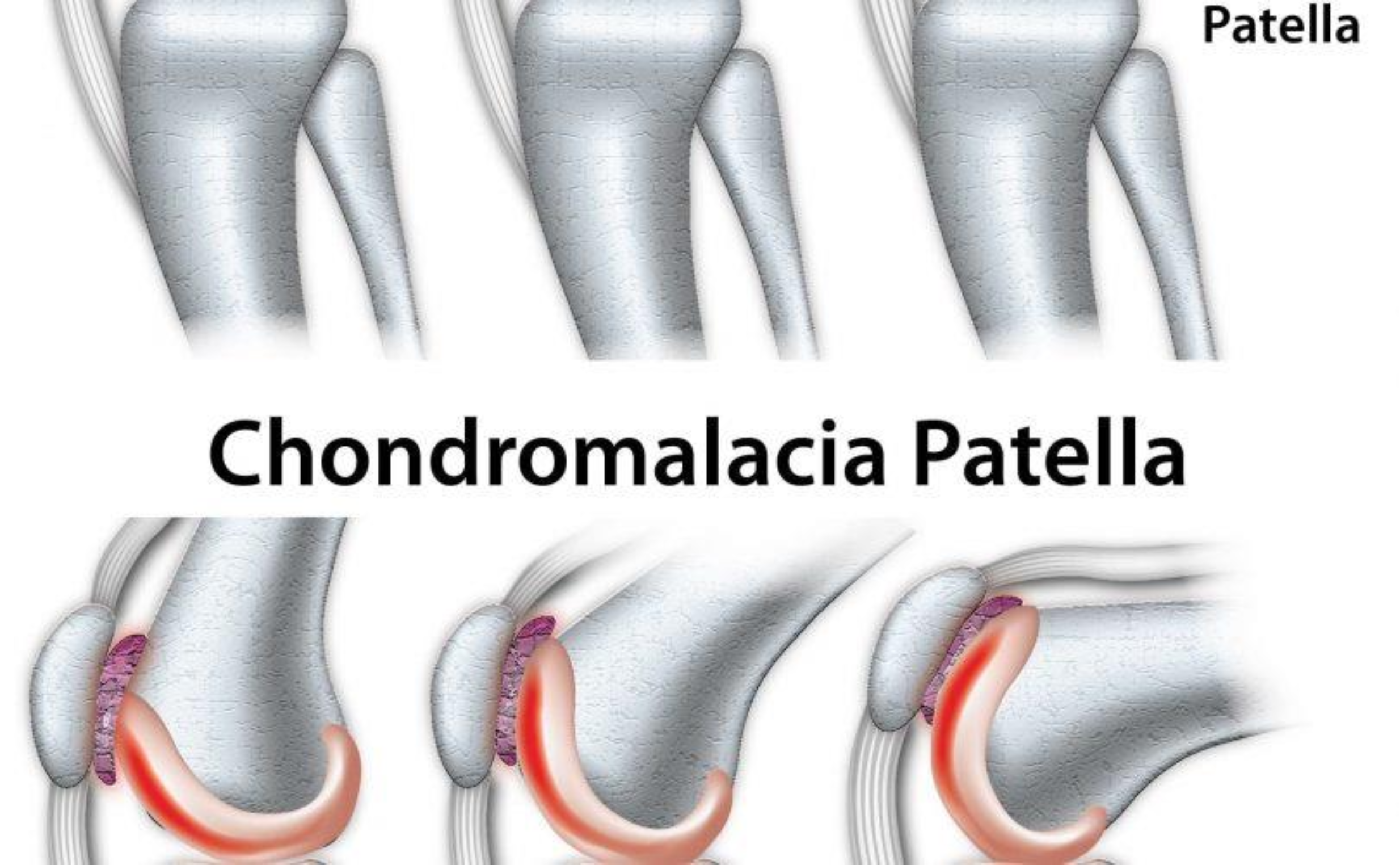
Chondromalacia patella (or patellae), also referred to as “runner’s knee” is a condition in which the cartilage cushioning the area under the patella (kneecap) begins to deteriorate and wear out. Due to this, the kneecap may start to rub against the femur (thigh bone) and cause discomfort or pain.
Causes of Chondromalacia Patellae
Osgood-Schlatter disease (OSD) is a growth-related, overuse injury most commonly seen in young adolescents. This disease is characterized by a painful inflammation (bump) located at your child’s tibial tuberosity. Your child’s tibial tuberosity is located about an inch below their kneecap (patella) at the growth plate where their patellar tendon attaches to their shinbone (tibia). OSD typically occurs during periods of rapid growth, when bones, muscles, and tendons are developing.
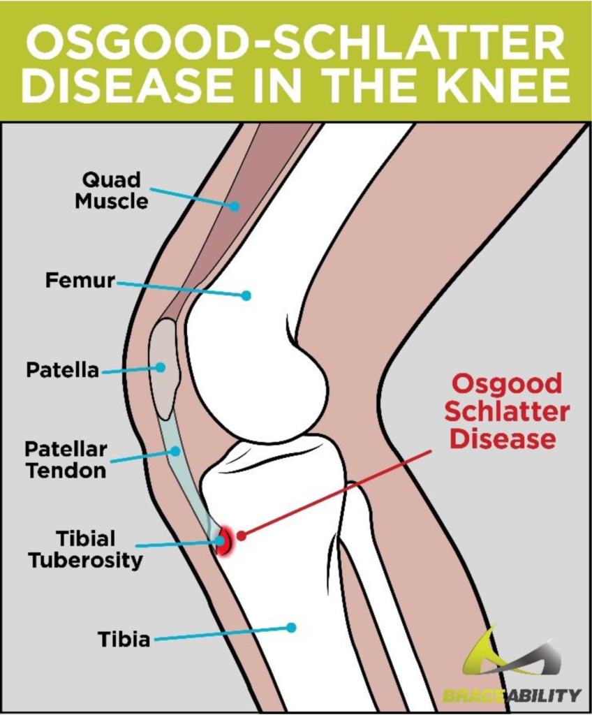
Signs and symptoms of Osgood-Schlatter
disease include:
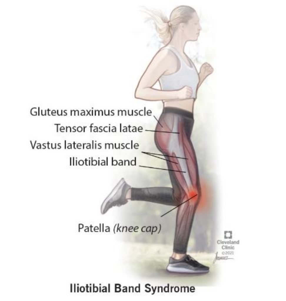
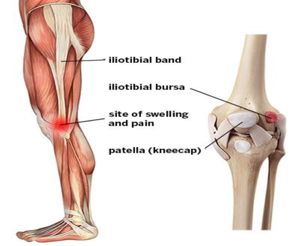
Causes of IT band syndrome.
ITBS is caused by excessive friction from the IT band being overly tight and rubbing against bone. It’s primarily an overuse injury from repetitive movements. ITBS causes friction, irritation, and pain when moving the knee. It seems to happen only in some people, though the reasons for this are unclear.
It’s especially common for cyclists and runners. It can even develop from repetitively walking up and down stairs, wearing high heels, or sitting for long periods with bent knees.
Symptoms of IT band syndrome may include:
How is iliotibial band syndrome treated?
Most cases of iliotibial band syndrome heal on their own without
surgery. An athlete may need to take time off from sports to give
their leg time to recover. The amount of time depends in part on the
extent of the injury.
Treatment may include:
The ACL is one of the major ligaments in the knee, responsible for providing stability and preventing excessive forward movement of the tibia in relation to the femur. They occur most frequently in those who play sports involving pivoting (e.g. football, basketball, netball, soccer, European team handball, gymnastics, downhill skiing). They can range from mild (such as small tears/sprain) to severe (when the ligament is completely torn).
Causes for ACL Tear
ANTERIOR CRUCIATE LIGAMENT [ ACL] injuries can occur due to various factors, including;
Diagnosis for ACL Tear
ACL injuries are usually diagnosed based on the combination of clinical examination, imaging studies [such as MRI], and
assessment of symptoms and mechanism of injury. Often the diagnosis can be made on the basis of the physical exam alone,but you may need tests to rule out other causes such as [pivot-shift test,anterior drawer test and lachman test] to determine the severity of the injury.
Treatment for ACL Tear
A medial collateral ligament [MCL] sprain is a common injury that affects the ligament on the inner side of the knee. The MCL play a crucial role in stabilizing the knee joint by preventing excessive side-to-side movement. A sprain occurs when the ligament is stretched beyond its normal range of motion or when it is torn partially or completely.
Common symptoms of an injury to the medial collateral ligament are:
Common Diagnosis of an injury to the medial collateral ligament are:
A doctor will typically diagnose an MCL sprain through a physical exam, which may include:
Common Treatment of an injury to the medial collateral ligament are:
A Meniscus tear is a common injury that involves damage to the cartilage disc [menisci] located between the thigh bone [femur] and shinbone [tibia]. These C-shaped discs act as a shock absorber and help stabilize the knee joint.
Causes of Meniscus Tear
Most often, the meniscus tears during a sudden motion in which your knee twists while your foot stays planted on the ground. The tear frequently occurs while playing sports. People whose cartilage wears down (due to age or arthritis) can tear a meniscus from a motion as simple as stepping on an uneven surface. Sometimes, degeneration from arthritis causes a tear.
Types of Meniscus Tears
Meniscus tears can be classified based on their location, severity, and pattern. Common types of meniscus tears include:
Diagnosis of Meniscus Tear
Treatment of Meniscus Tear
The treatment of a meniscus tear depends on several factors including the severity of the tear, the patient’s age, activity level, and overall health. Here are some common treatment options:
Conservative Treatment:
Injection Therapy:
Surgical Treatment:
Rehabilitation:
Kneecap dislocation occurs when the round-shaped bone covering the knee (patella) moves or slides out of place. The dislocation often occurs toward the outside of the leg.
Causes of Patellar Dislocation
Symptoms of Patellar Dislocation
Typical symptoms include:
How is Patella Dislocation Diagnosed?
Healthcare providers can usually diagnose a dislocated kneecap by physically examining the knee and asking you questions about the injury. However, they’ll order radiographic imaging tests to check for any related injuries, such as torn ligaments, cartilage injury or fractures. With patellar dislocation, it is safe to correct the joint first and take pictures after.
If your dislocated patella corrected itself, you might not realize that it was dislocated. A dislocation that corrects itself is called “transient.” Afterward, your knee will still be sore and swollen, but it may look like many other more common knee injuries. In this case, imaging tests can show evidence after the fact that there was a dislocation, along with secondary injuries.
Treatment of Patellar Dislocation
Surgery:
An LCL injury is a sprain or tear to the lateral collateral ligament (LCL). The LCL is a band of tissue on the outside of your knee. It connects your thighbone to the bone of your lower leg and helps keep the knee from bending outward.
Causes of LCL Tear
LCL tears typically happen when you’re playing a sport that involves:
Diagnosis of LCL Tear
Treatment of LCL Tear
Treatment for a lateral collateral ligament (LCL) tear typically depends on the severity of the injury. Here’s an overview of potential treatment options:
Please complete the form below to request an appointment. A member of our intake team will follow up with you.
Vraj Complex, 302, 100 Feet Rd, Near Shyamal Cross road, Jodhpur Village, Ahmedabad, Gujarat 380015